Research
Research
Welcome to the Kasza Living Materials Lab at Columbia University. We are an interdisciplinary group of engineers, biologists, and physicists that work to understand how cells self-organize to build tissues with mechanical and structural properties that are required for proper function. We study this problem in the context of embryonic development to uncover fundamental mechanisms of how cells build tissues in vivo, and we use the fruit fly as a model organism. The goal is to use this fundamental understanding both to shed light on human health and disease and also to learn how to better build tissues in the lab.
Mechanical cues coordinating cell behaviors during morphogenesis
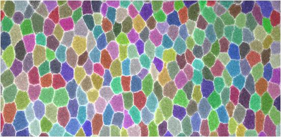
During embryonic development, groups of cells reorganize and build functional tissues with complex form and structure. In Drosophila, the coordinated actions of thousands of cells moving together rapidly narrow and elongate the embryo, doubling the length of the head-to-tail body axis. While much is known about the molecules required for these movements, less is understood about how cells work together (instead of against each other) to produce macroscopic changes in tissue shape and structure. Several lines of evidence point toward mechanical cues as key factors for coordination. This project employs high-resolution live confocal imaging, biophysical studies, and optogenetics to understand how mechanical cues coordinate cell movements during development.

Biophysics of cell rearrangements and tissue flows
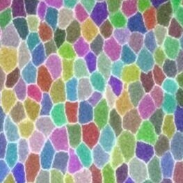
Tissue reorganization during development can be rapid and dramatic, often occurring through embryo-scale flows involving thousands of cells. For example, the epithelium in the Drosophila embryo doubles its length in just 30 minutes. This occurs primarily through cell rearrangements, in which cells change their relative positions within the tissue. This type of tissue movement or flow is highly conserved, and can be driven by internal forces generated by cells or by external forces. It is not yet clear how cell rearrangements and tissue fluidity are regulated. In this project, live imaging, genetics, and biophysical approaches are combined to dissect how cell properties and interactions are regulated to allow (or prevent) rapid cell rearrangements and tissue flows during developmental events.

Optogenetic control of forces In MuLTICELLULAR TISSUES
Emerging optogenetic technologies allow unprecedented control over protein activities inside cells with high spatiotemporal precision. We are using optogenetics to control the forces generated by cells within developing embryos. This project employs optogenetics to understand how mechanical forces bend, fold, and stretch tissues and also feedback on biochemical events to control cell behavior and cell fate. We are also using these tools to build and shape cultured tissues in the lab with desired shapes and structures.